Signs & Symptoms
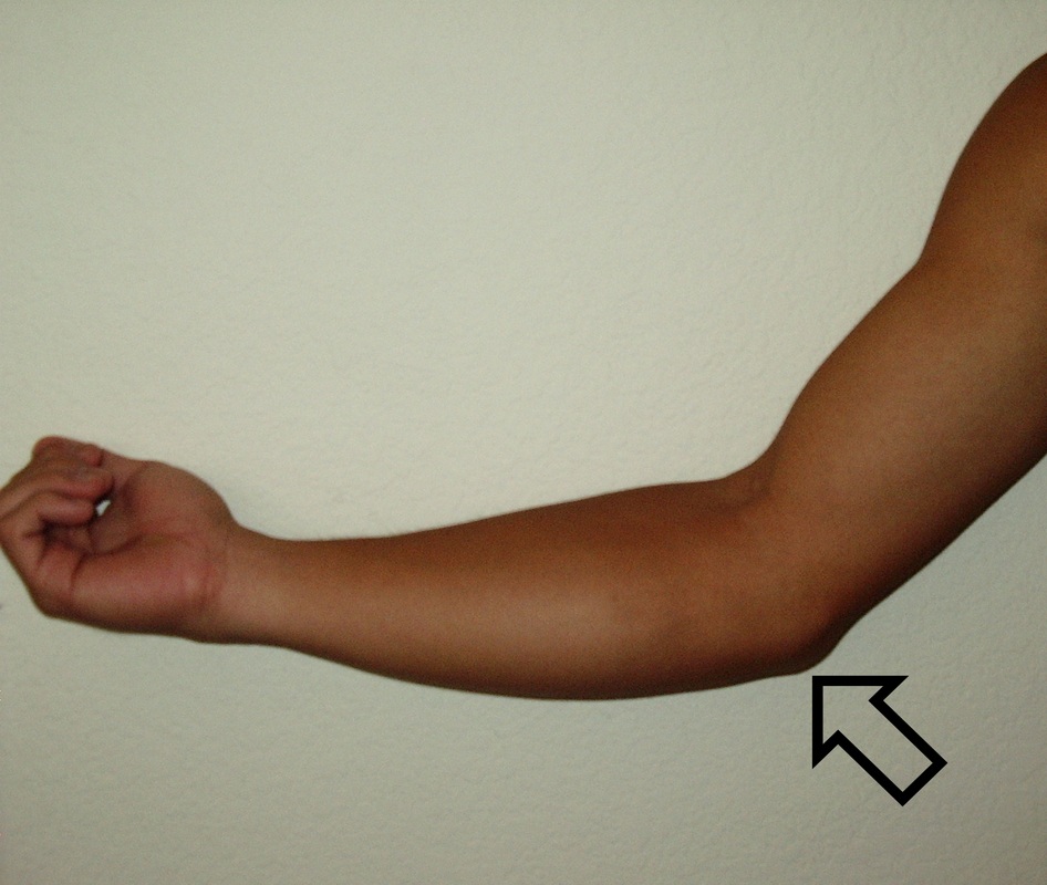
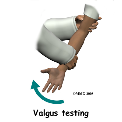
The two most common symptoms are tenderness and swelling in the medial elbow, specifically the distal attachment of the ulnar collateral ligament on the ulna. The athlete may report hearing a pop or pulling sensation followed by a loss of ability to grip strongly. Palpation may reveal crepitus from the medial epicondyle to the coronoid process. Reduced range of motion may follow, especially an inability to fully extend elbow and flex the wrist. A positive valgus elbow stress test is common. This special test needs to be done with 20-30° of elbow flexion aiming to find an end-point. Another test that may be positive is the Tinel sign, which would indicate injury to the ulnar nerve or elbow subluxation. Functional tests will reveal a significant decrease in throwing velocity.
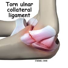
A Magnetic Resonance Imaging (MRI) can complete full diagnosis. This will determine the extent of the UCL sprain; with a grade III sprain a complete ligament rupture will be found. An x-ray may be advised by physicians to check the integrity of the humeral condyle and olecranon. It also would reveal loose bodies in the elbow joint or calcification that may have lead to the sprain. After diagnosing this pathology with the imaging equipment, surgery should be initiated as soon as acute swelling and inflammation have become manageable for the surgeon.
Valgus Stress Test Instructional Video
The two most common symptoms are tenderness and swelling in the medial elbow, specifically the distal attachment of the ulnar collateral ligament on the ulna. The athlete may report hearing a pop or pulling sensation followed by a loss of ability to grip strongly. Palpation may reveal crepitus from the medial epicondyle to the coronoid process. Reduced range of motion may follow, especially an inability to fully extend elbow and flex the wrist. A positive valgus elbow stress test is common. This special test needs to be done with 20-30° of elbow flexion aiming to find an end-point. Another test that may be positive is the Tinel sign, which would indicate injury to the ulnar nerve or elbow subluxation. Functional tests will reveal a significant decrease in throwing velocity.
A Magnetic Resonance Imaging (MRI) can complete full diagnosis. This will determine the extent of the UCL sprain; with a grade III sprain a complete ligament rupture will be found. An x-ray may be advised by physicians to check the integrity of the humeral condyle and olecranon. It also would reveal loose bodies in the elbow joint or calcification that may have lead to the sprain. After diagnosing this pathology with the imaging equipment, surgery should be initiated as soon as acute swelling and inflammation have become manageable for the surgeon.
Valgus Stress Test Instructional Video
